The Brain's Hidden Supply Line: How Blood Proteins Could Change Alzheimer's Research
Scientists have identified 92 blood proteins that cross into the brain. A new mouse study reveals how this transport system may be disrupted in early Alzheimer's.
The Brain’s Hidden Supply Line: How Blood Proteins Could Change Alzheimer’s Research
The blood–brain barrier is usually described as the brain’s protective wall. New research suggests it also operates as a carefully controlled delivery system, importing proteins that help maintain the cells responsible for healthy brain function.
For decades, the blood–brain barrier has been understood primarily as a defense mechanism. It separates circulating blood from the delicate environment of the brain, restricting the entry of potentially harmful substances while allowing essential nutrients and selected molecules to pass.
But what if this barrier does more than keep unwanted materials out?
A study published in Cell on November 11, 2026, offers a more detailed picture of the relationship between the bloodstream and the brain. Researchers identified dozens of proteins that can cross the barrier and reach specific types of brain cells. They also found that the movement of some of these proteins was disrupted in mice with Alzheimer’s-like disease.
The finding points toward a potentially important connection between organs outside the brain and the biological systems that help preserve it.
It does not establish a new Alzheimer’s treatment, and the experiments were conducted in mice. Nevertheless, the work raises a significant research question: could some early neurological problems involve the failure of the brain to receive proteins that it normally depends on?
The blood–brain barrier is more than a wall
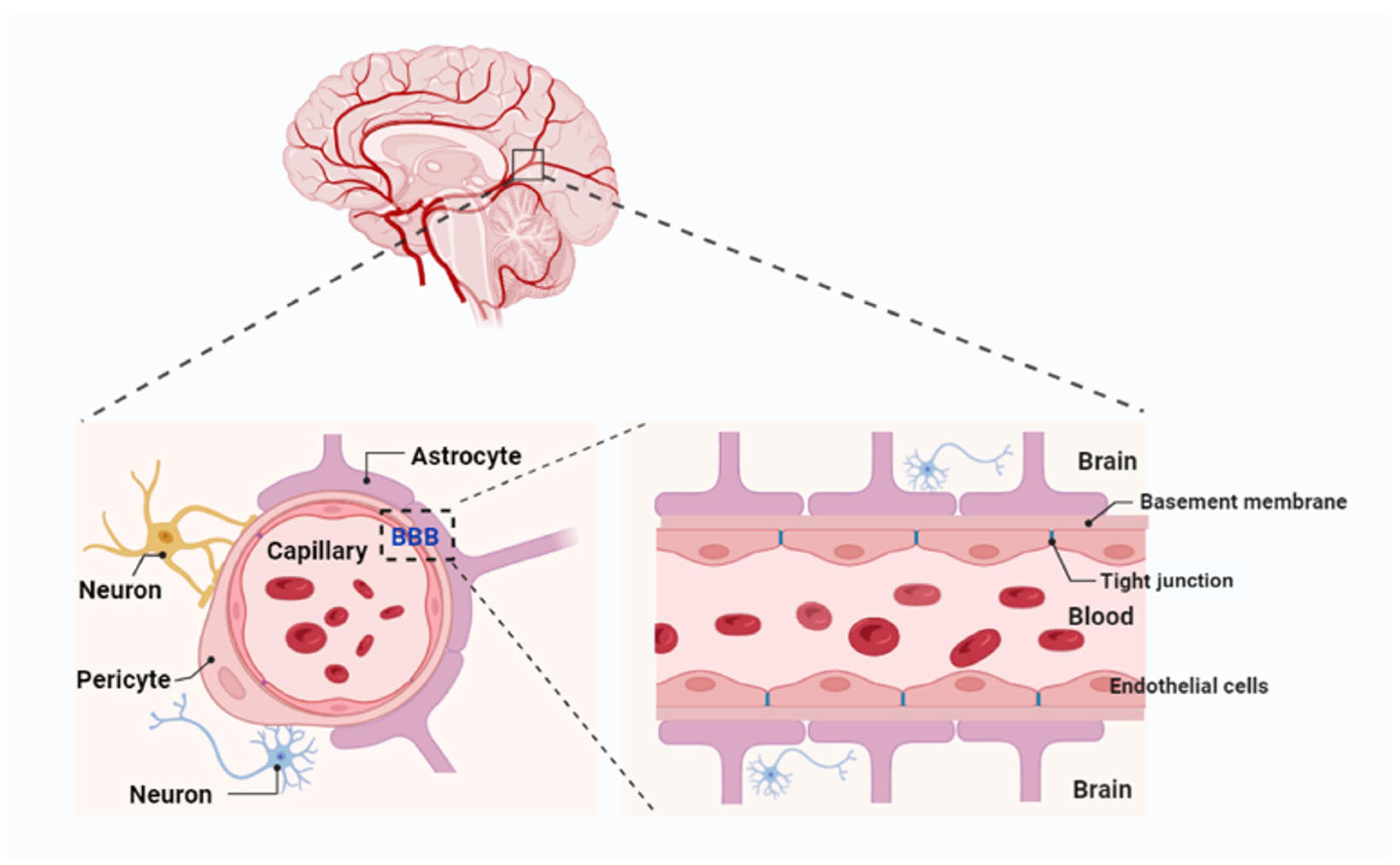
The blood–brain barrier is formed largely by specialized endothelial cells lining the brain’s smallest blood vessels. These cells are connected by tight junctions that restrict the movement of substances between the bloodstream and brain tissue.
Other components, including pericytes and astrocytes, help regulate the barrier’s function.
This arrangement is essential because the brain operates within a tightly controlled chemical environment. Sudden changes in circulating substances could interfere with the electrical and biochemical processes that allow neurons to communicate.
However, the barrier is not completely sealed.
Glucose, amino acids and other essential substances cross through specialized transport mechanisms. Certain proteins can also move across through processes involving receptors and cellular transport.
The new research examines this less familiar side of the system: the selective movement of proteins from the blood into the brain.
Rather than treating the barrier as a passive filter, the researchers describe it as an active interface that can recognize, transport and direct specific molecules.
Researchers tracked 861 proteins
The research team developed a method using bioorthogonal proteomics to label circulating proteins and follow their movement into brain tissue.
They examined 861 blood proteins in their experimental system. Of these, 92 were detected crossing the blood–brain barrier and entering the brain parenchyma.
The researchers also identified a broader set of 191 plasma proteins sampled by the barrier, helping them investigate which circulating molecules were being handled by this interface.
Many of the proteins that reached the brain were associated with carrier and metabolic functions. Several originated in organs outside the nervous system, particularly the liver.
This is an important detail.
The brain is often discussed as though it operates independently from the rest of the body. In reality, its cells depend on a continuous exchange of signals, nutrients and molecular resources.
The findings suggest that the liver and other organs may contribute to brain maintenance through proteins that travel through the bloodstream and are selectively delivered to particular brain cells.
The study also identified different destinations for different proteins. Some were associated with astrocytes, while others were directed toward microglia.
This suggests that protein transport is not simply a matter of allowing molecules into the brain. The destination of each molecule may also matter.
The liver protein that caught the researchers’ attention
One protein became particularly important in the investigation: apolipoprotein A1, commonly known as APOA1.
APOA1 is best known for its role in lipid transport and is a major component of high-density lipoprotein particles in the bloodstream.
The researchers found that APOA1 could cross the blood–brain barrier through a pathway involving the receptor SR-B1. Once inside the brain, it interacted with astrocytes through another protein, ABCA1.
Astrocytes are support cells with responsibilities extending far beyond their traditional description as structural helpers. They regulate the environment surrounding neurons, contribute to metabolic support and help maintain the connections through which brain cells communicate.
One important function of ABCA1 is regulating cholesterol movement out of cells.
Cholesterol is essential for neuronal function. It contributes to the structure of cell membranes and supports the formation and maintenance of synapses, the communication points connecting neurons.
The research therefore connects several biological processes:
- APOA1 originates in the circulation, including production in the liver.
- SR-B1 helps facilitate its passage across the blood–brain barrier.
- APOA1 interacts with ABCA1 in astrocytes.
- This interaction supports cholesterol movement associated with synaptic maintenance.
The significance is not that scientists have discovered a single protein capable of preventing Alzheimer’s disease. Rather, the study identifies a transport pathway that may help explain how the body’s metabolic systems contribute to the maintenance of brain connections.
What changed in the Alzheimer’s mouse models?
The researchers next examined mice engineered to develop features associated with Alzheimer’s disease.
They used an early-stage 5xFAD mouse model, which is commonly employed to investigate biological processes associated with amyloid-related pathology.
The results suggested that the movement of protective proteins across the blood–brain barrier was altered before the barrier showed the kind of broad physical leakage often associated with more advanced neurological damage.
In particular, the researchers observed reduced APOA1 uptake and increased uptake of another protein, SERPINA1, in their experimental model.
This is an important distinction.
The familiar description of Alzheimer’s-related blood–brain barrier dysfunction often focuses on the barrier becoming excessively permeable, allowing unwanted substances to enter brain tissue.
The new findings suggest that selective transport can also become disrupted. In other words, the problem may not only involve harmful substances getting through. It may also involve beneficial proteins failing to reach the cells that need them.
The study linked this altered transport with changes in astrocytes, reduced synaptic integrity and behavioral deficits in the mice.
It provides evidence for a different way of thinking about early communication between the bloodstream and the brain.
Could restoring APOA1 make a difference?
To investigate whether the disrupted pathway could be modified, the researchers increased APOA1 production in the liver of mice with Alzheimer’s-like disease.
The objective was to raise the amount of APOA1 available in the circulation and determine whether more of the protein could reach the brain.
The engineered mice showed improvements in several measured outcomes, including astrocyte-related changes, synaptic measures and behavioral deficits.
The researchers also reported reductions in certain disease-associated markers, including fat accumulation in astrocytes and inflammatory signals.
These results make the APOA1 pathway interesting as a potential research target.
However, an improvement in an engineered mouse model is not evidence that the same intervention will work in people.
The experimental approach involved deliberately changing protein production in animals. It does not establish that increasing APOA1 in humans would be safe, effective or capable of preventing or reversing Alzheimer’s disease.
Further work is needed to determine how the pathway behaves in human biology and whether it can be modified without creating unwanted effects elsewhere in the body.
A different way to think about brain health
The study contributes to a growing body of research examining how the brain communicates with other organs.
Scientists increasingly investigate the connections between the brain, liver, immune system, gut and cardiovascular system. These relationships are sometimes discussed under the broader concept of the body–brain axis.
The new findings add a specific molecular mechanism to that discussion.
If circulating proteins can be selectively delivered to particular brain cells, then changes in the production, availability or transport of those proteins could influence neurological function.
This may also help researchers investigate why some neurological conditions involve changes in the brain’s supporting cells before widespread neuronal damage becomes apparent.
Astrocytes and microglia are particularly important in this context. They help regulate the environment in which neurons operate, and changes in their behavior can influence inflammation, metabolism and synaptic function.
The research does not suggest that the liver alone determines brain health. It instead demonstrates one pathway through which a distant organ can contribute to processes inside the brain.
What the discovery does not prove
The distinction between a promising biological mechanism and a medical breakthrough is essential.
The research was conducted in mice, including an engineered model of Alzheimer’s disease. Mouse models are valuable for investigating disease mechanisms, but they do not reproduce every aspect of human Alzheimer’s disease.
Several questions remain unanswered.
First, researchers need to establish whether the same APOA1 transport changes occur in people during the earliest stages of Alzheimer’s disease.
Second, it remains necessary to determine whether altered protein transport contributes directly to disease progression or partly reflects other biological changes already taking place.
Third, any attempt to modify APOA1 production or transport would need careful evaluation of safety, dosage, long-term effects and interactions with other metabolic systems.
Finally, identifying 92 proteins that can cross the barrier does not mean that all of them have established protective effects. Their individual functions, destinations and contributions to brain health require further investigation.
The study is therefore best understood as a new map of a biological transport system, not as a treatment announcement.
The brain may depend on a wider network than previously understood
The blood–brain barrier has long represented one of neuroscience’s most important protective systems. Its ability to restrict harmful substances is fundamental to the brain’s survival.
But protection is only part of its role.
The new research suggests that the barrier also helps coordinate a selective supply network, bringing proteins from the bloodstream into the brain and directing them toward particular cell types.
That possibility broadens the way scientists think about neurological health. Brain function may depend not only on what happens inside the skull, but also on the molecular exchanges that connect the brain with organs throughout the body.
For Alzheimer’s research, the next step is to determine whether this transport system can be measured and understood in humans, and whether its disruption offers a meaningful opportunity for future intervention.
The most important insight may be a simple one: the brain is not an isolated organ. Its health is partly shaped by the messages and materials it receives from the rest of the body.
Sources and Further Reading
- Zhou, H. et al. (2026). “The blood-brain barrier imports diverse neuroprotective proteins through pathways impaired early in Alzheimer’s disease models.” Cell. DOI: 10.1016/j.cell.2026.10.039.
- Basu, M. (2026). “New culprit in early Alzheimer’s: blood–brain barrier blocks protective proteins.” Nature, November 11, 2026. Read the report.
- Reuters (2026). “Blood-brain barrier discovery could lead to new Alzheimer’s disease approach.” November 13, 2026. Read the report.
- Gladstone Institutes — Research institution associated with the study. Visit Gladstone Institutes.
Research note: The principal experimental findings discussed here come from animal models. They should not be interpreted as evidence of a proven human treatment or prevention strategy.